The Greatest Guide To Ultrasound Scan - Tests And Scans
This innovation can assist detect and deal with specific conditions - diagnostic ultrasound. Ultrasound imaging has many usages in medication, from confirming and dating a pregnancy to detecting certain conditions and directing medical professionals through precise medical procedures. Ultrasound images have numerous uses throughout pregnancy. Early on, they may be used to determine due dates, reveal the existence of twins or other multiples, and rule out ectopic pregnancies.
Numerous expectant moms and dads look forward to finding out the sex of their children via ultrasound midway through a pregnancy. And later in pregnancy, doctors can even use ultrasounds to approximate how large a child is right before shipment. Medical professionals employ ultrasound imaging in diagnosing a variety of conditions impacting the organs and soft tissues of the body, including the heart and blood vessels, liver, gallbladder, spleen, pancreas, kidneys, bladder, uterus, ovaries, eyes, thyroid, and testicles.
The Buzz on Medical Ultrasound
Ultrasound imaging can help doctors throughout procedures such as needle biopsies, which need the physician to eliminate tissue from an extremely accurate area inside the body for testing in a lab. Ultrasounds sometimes are used to find and deal with soft-tissue injuries. The majority of ultrasounds are done using a transducer on the surface area of the skin.
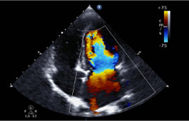
A transrectal ultrasound is often utilized in the diagnosis of prostate conditions. A transesophageal echocardiogram uses the transducer probe in the esophagus so that the sonographer can acquire clearer images of the heart. Furthermore, ultrasound innovation has actually advanced to enable various kinds of imaging: Doppler is an unique kind of ultrasound that creates images of blood flow through vessels.
Some Ideas on What Else Is An Ultrasound Used For? You Need To Know
Echocardiograms are utilized to see the heart. 3D imaging adds another measurement to the ultrasound image, creating three-dimensional interpretations rather than the flat two-dimensional images that are made with conventional ultrasound. 4D ultrasounds program 3D images in motion. Ultrasounds use many advantages: They are generally pain-free and do not need needles, injections, or incisions.
In fact, there are no recognized damaging impacts when used as directed by your healthcare provider. Ultrasound records images of soft tissues that do not show up well on X-rays. Ultrasounds are widely available and less costly than other approaches. Depending on the kind of ultrasound test you are having, your medical professional may offer unique guidelines, such as not consuming or drinking anything for a number of hours prior to the test.
What Does Ultrasound Scans: How Do They Work? Do?
You should wear comfortable clothing that is easy to remove or partly remove. In some cases, you might need to disrobe or use a dress, but frequently an ultrasound technician can easily access the location of the body that is being evaluated without your having to take off your clothes.
This is so the transducer can quickly move throughout your skin without any air in between. They may be looking for specific markers and may make measurements or notes while the test remains in development. A typical ultrasound takes in between 30 minutes and an hour. Ultrasounds usually are not unpleasant, and you are awake and alert throughout the treatment.
The 3-Minute Rule for Ultrasonography And Pelvic Ultrasound
SOURCES: American Society of Radiologic Technologists: "Ultrasound." FDA Customer Health Info: "Taking a Close Appearance at Ultrasound." Mayo Center. RadiologyInfo. org: "General Ultrasound Imaging." 2019 WebMD, LLC. All rights reserved.
Existing as of: December 8, 2019 Author: Medical Review:Kathleen Romito MD - click site Family Medicine & Adam Husney MD - Household Medicine & Martin J. Gabica MD - Household Medicine & Howard Schaff MD - Diagnostic Radiology.
Some Known Questions About Chest Ultrasound.
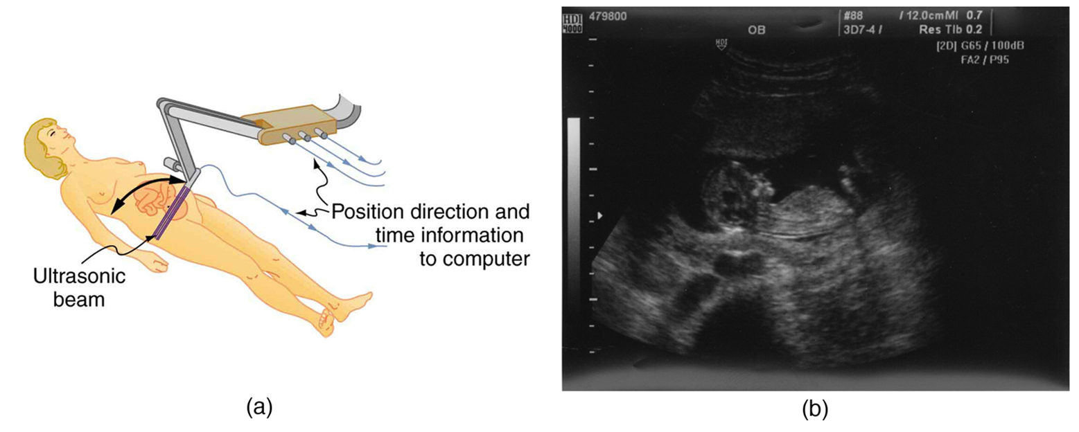 ultrasound; scan; ultra; sound; waves; placenta; several; Contents Ultrasound utilizes acoustic waves to produce an image (image). The acoustic waves can not be heard and the power of the sound waves used is very low. In pregnancy an ultrasound scan can be used to look at the establishing infant, the uterus and the placenta.
No proof has been found of ultrasound triggering any harm to either the mother or establishing child. In most industrialized countries about 90% of ladies have at least one ultrasound throughout their pregnancy. Pregnancy is an amazing time for both parents, but there can be concerns. Speak with your physician or midwife if there are any concerns or questions about your pregnancy and for more info before you choose whether to have a test done.
The Only Guide for Abdominal Ultrasounds: Purpose, Procedure
The most common ones are: To determine viability of pregnancy (check if the infant is alive) To inspect the variety of children present To work out the diagnostic ultrasound surrey age of child in a dating scan To inspect the infant's development and physical advancement To monitor the pregnancy if there have been complications, such as bleeding, fluid loss, high blood pressure (hypertension) or gestational diabetes To inspect the position of the placenta and assess quantity of amniotic fluid around the baby To keep an eye on placental health and function.
This is the finest time to examine the infant's physical development. Ultrasound scans can be done at any stage of pregnancy, with indications of pregnancy being viewed as early as 5 weeks. They are utilized to offer different info at different times throughout the pregnancy. Ultrasound is an outpatient treatment (you will not be admitted to health center), and it is done by a specially trained and certified sonographer or medical professional.
Our Abdominal Ultrasounds: Purpose, Procedure Ideas
The length of the scan will depend on the factor for the scan and can take from hr to about 1 hour or more. Your partner or a support individual will generally have the ability to be with you during the scan. Sometimes, due to the position of the infant, excellent views can not be gotten and a repeat scan is needed at a later date.
If you do not wish to view the screen, inform the sonographer before the scan starts. For ultrasounds early in pregnancy the finest views are gotten if the woman's bladder is complete. The uterus is typically hidden behind the bowel making it tough to see. When the bladder is full the bowel is pushed out of the way - private ultrasound.